


English
Request a Quote


The appearance of the solution is a must test item for raw material used for injection, which usually includes the clarity, turbidity and color of liquid. In the manufacturing of biologics, pigments are found in protein intermediates and final sample stock solutions due to contact between the medium and metal processing equipment. This issue is particularly noticeable in highly concentrated protein solutions.
The appearance of the protein solution is an important quality parameter in product release testing. The European Pharmacopoeia sets requirement on color of monoclonal antibodies for human use, i.e., liquid formulations of monoclonal antibodies for human use shall be "clear or slightly milky, colorless or slightly colored liquids" [1].
This is also a test item specified in The International Council for Harmonisation of Technical Requirements for Pharmaceuticals for Human Use (ICH) Q6B releases specification. Abnormal color means the possibility of contamination or degradation of product, making the control and removal of pigments an essential part of protein purification process. A good understanding of pigments control and removal can be very helpful in drafting and filing quality standards for new or generic drugs.
Pigments which affect protein solution color and are tricky to remove are produced when the medium components bind to or denature protein molecules during culture and harvesting. The most influential pigments are Vitamin B and iron ions. For example, CN-B12, which is usually added to mammalian cell culture medium, will convert to OH-B12 under exposure to light and become pink after binding with antibody molecules. The increase of iron ions concentration will darken the colour of protein solution and raise the level of acid variant.
Iron ion does not directly cause changes in protein solution, but produces reactive oxygen species (ROS) due to Fenton reaction, which oxidizes proteins, resulting in the oxidation of tryptophan in proteins to N-formylkynuanine (light yellow) and kynuanine (brown yellow)[3,5].
Several advanced glycation end products (Ages) produced during cell culture procedure are colored (yellowish brown) and fluorescent. The structure of AGE and its formation are complex and diverse, but they all stem from the amino side chain reaction between the dicarbonyl byproducts of oxidative degradation of reducing sugars or carbohydrates and protein lysine or arginine [3].
In the middle and late stage of Pichia pastoris induction, AOX1 alcohol oxidase is released into the fermentation solution and binds to flavin adenine dinucleotide (FAD) cofactor to form an octomer, resulting in green color [4].
In addition, natural interacting proteins and small molecular pigments may also introduce color.
During the protein production process, effective control and removal of pigments can be conducted from cell line construction, upstream culture and chromatographic purification. Especially in the chromatographic purification stage, where resins can be used for the removal of pigments.
● Cell line construction stage
Different cell lines have various adsorption and demand for iron ions and Vitamin B12. To reduce the possibility of abnormal color to very low level, it is recommended to choose cell lines having fast consumption rate or low demand for iron ions and Vitamin B12, which will reduce the level of iron ions and Vitamin B12 in culture medium.
● Upstream culture
Optimize culture medium components. For example, adjust the content of highly oxidizing components (e.g. iron ions) and light sensitive components (e.g.B Vitamins.etc.) in the culture medium during antibody production. In the process development of cell culture, optimizing metal ions or content of other additives might cause coloring of protein. It is recommended to try different types of culture media to reduce the consumption of these substances. Meanwhile, high-yield cell line screening or long-term domestication can help to ensure productivity.
● Chromatographic separation stage
Pigments can take in various types, specifically, macro-molecule pigment, micro-molecule pigment, pigments with positive/negative charges, hydrophobic and hydrophilic pigments. Chromatographic separation is used for the removal of pigments with no binding or non-covalent bonding to proteins. For some pigments which will covalent bind to proteins (e.g bind between OH-B12 and cysteine residues), it is necessary to optimize culture condition and control condition in upstream culture stage. In case of unfamiliarity with pigments property in solution, it is possible to try different chromatographic methods.
1. Affinity chromatography
Affinity chromatography is usually applied in the capture stage in protein purification. Pigment can be effectively removed at the same process of protein accumulation by damaging ionic adsorption and non-specific adsorption between pigment and resin via optimized buffer system.
2. Ionic exchange chromatography
In neutral condition, most pigments carry negative charges. Under anion exchange flow-through mode, pigment can bind with resin and then being eluted by high salinity or alkali; alternatively, target protein can bind with cation exchange resin while undesirable pigments flow through. Solution pH and ionic strength play vital roles in the purification process, which means suitable sample loading condition is the key for effective removal of pigment.
3. Hydrophobic interaction chromatography(HIC)
Pigment has certain level of hydrophobicity, which can be removed by using the surface hydrophobic disparity in pigment and target protein. Specifically, pigment removal methods include resin type optimization, salt type and concentration selection, gradient elution method.
4. Size exclusion chromatography(SEC)
In case of significant molecular weight disparity between pigment and target protein, SEC resin can be used for the removal of pigment. For instance, in the process of buffer exchange by desalting column or impurities removal via high resolution resin in polishing step, pigment can be removed or adsorbed to certain extend.
Based on the DoE developed method, Diamond MMC Mustang resin was used in the purification process to remove pigment from in vitro secretory enzymes of animal origin. The resin did a great job in the removal of yellow pigment. As illustrated in the following picture, eluent was almost colorless, indicting good effect to pigment removal.
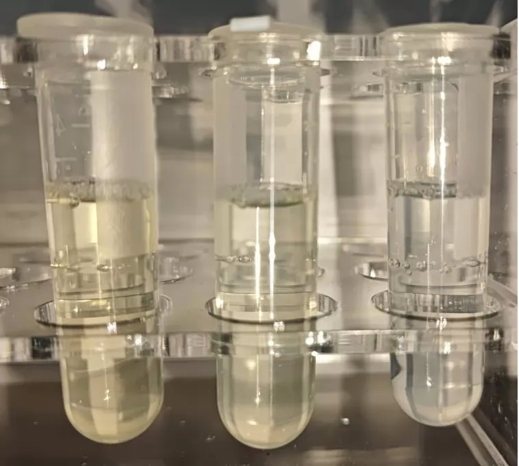
Left: Original sample
Middle: Washing buffer
Right: eluent
Diamond MMC Mustang resin is combination between high rigid weak AEX resin and hydrophobic Mixed-mode resin. Due to its IEX, HIC, hydrogen bond and van der Waals force function, the resin enjoys good performance in the removal of various pigments.
Pigments possess complex properties. It is ideal to design CIP plan according to the tolerance of resins. NaOH is the most commonly used reagent for cleaning. Apart from that, high salinity solution, organic reagents (20% ethanol or 30% isopropanol), urea, GuHCl or acids are also widely chosen for cleaning approach. It is also possible to process resin with mixed reagents or other more targeted cleaning methods. For example, choose specific cleaning method according to the property of pigment.
● The removal of Iron ions residue-caused yellow pigment
The yellow pigment is mainly from Fe(OH)₃ precipitation formed by chemical reaction between iron ions and NaOH. It is possible to try washing with acids. For example, process the sample with 0.5M H₃PO₄ for 10-15min. Please notice that the processing time shall not be too long, which might cause re-emergence of iron ions binding.
● The removal of Vitamin B12-caused pink pigment
It is possible to replace OH radicals in OH-B12 with electron donors such as histidine or imidazole. For example, wash wish 5-10CV of 0.3M histindine+0.5M NaCl buffer at pH 4.0.
● The removal of hydrophobic interaction bond pigments
To use a high concentration of alkaline solution to destroys the protein structure and hydrophobic force. For example, process with 0.5~1M NaOH for 3-5CV.
● The removal of lipid pigment
According to the principle of similarity and dissolution, it is possible to process samples with 3-5CV of nonionic detergent, 70% ethanol, 30% isopropanol.
● The removal of deposited stubborn pigment
It is possible to process with multiple types of reagents. For example, process with 0.5M CH₃COOH + 8M urea+ 1-2M NaCl or NaOH+2M NaCl for 5-10CV. For samples can tolerate organic solvents, it is possible to wash with 1M NaOH+30% isopropanol.
As the resin using number increases, accumulated pigments might cause darkening of resin color, decreased binding capacity, even resin bed collapse, resin hardening, compromising resin performance and life cycle. Thus, it is essential to remove pigments in resin.
Pigments enjoy great diversity. Despite being one of the most effective methods for pigment removal, chromatography resin performance will be impacted by pigment property. Thus, filtration and clarification in the pre-processing of feedstock can dramatically mitigate purification pressure in subsequent steps as well as extend life cycle of resins.
Reference
[1] European Pharmacopoeia, Council of Europe, Strasbourg,France, Edition 7.5; 2012.
[2] Kenneth M Prentice, et al. Hydroxocobalamin association during cell culture results in pink therapeutic proteins [J]. mAbs, 5:6, 974-981, 2013.
[3] Margaret Butko, et al. Recombinant Antibody Color Resulting from Advanced Glycation End Product Modifications [J]. Analytical Chemistry, 86(19): 9816-9823, 2014.
[4] L. M. Damasceno, et al. An optimized fermentation process for high-level production of a single-chain Fv antibody fragment in Pichia pastoris [J]. Protein Expression and Purification, 37:18-26, 2004.
[5] Weng Zhi-Bing, Wang Peng-Hong, ZHAO Li, et al. A new method for the removal of stubborn pigments from chromatographic fillers [J]. Chinese Journal of Food and Biotechnology,2022,41(02):106-111.

Jun
26

Jun
12
May
21
.png)


.png)
.png)
.png)

.png)