


English
Request a Quote


Efficient production of antibody therapeutics relies on a precise and robust downstream purification process. It serves as the critical bridge between upstream expression and final drug products, and is a central control point for both product quality and manufacturing cost. This article provides a systematic overview of the fundamentals of antibody therapeutics and offers an in-depth analysis of key downstream purification technologies, delivering a comprehensive technical reference for professionals engaged in antibody development and manufacturing.
Antibody purification is a biotechnological process used to selectively isolate, concentrate, and purify antibody proteins from complex biological samples such as serum, cell culture supernatants, and ascites fluid, based on differences in physicochemical properties and molecular structure.
The primary objective is to remove impurities including host cell proteins, salts, and cell debris, thereby obtaining antibodies with high purity, structural integrity, and preserved biological activity. This process is essential in antibody preparation, immunoassays, antibody drug development, and basic biomedical research.
Before discussing purification methods and workflows, it is important to understand the structure and classification of antibodies.
Antibodies, also known as immunoglobulins (Ig), are key effector molecules of the immune system responsible for recognizing and neutralizing foreign antigens such as bacteria and viruses.
Structurally, all antibody monomers adopt a “Y” shape and are composed of four polypeptide chains: two identical heavy chains (H chains) and two identical light chains (L chains). These chains are linked by disulfide bonds to form a symmetric tetrameric structure.
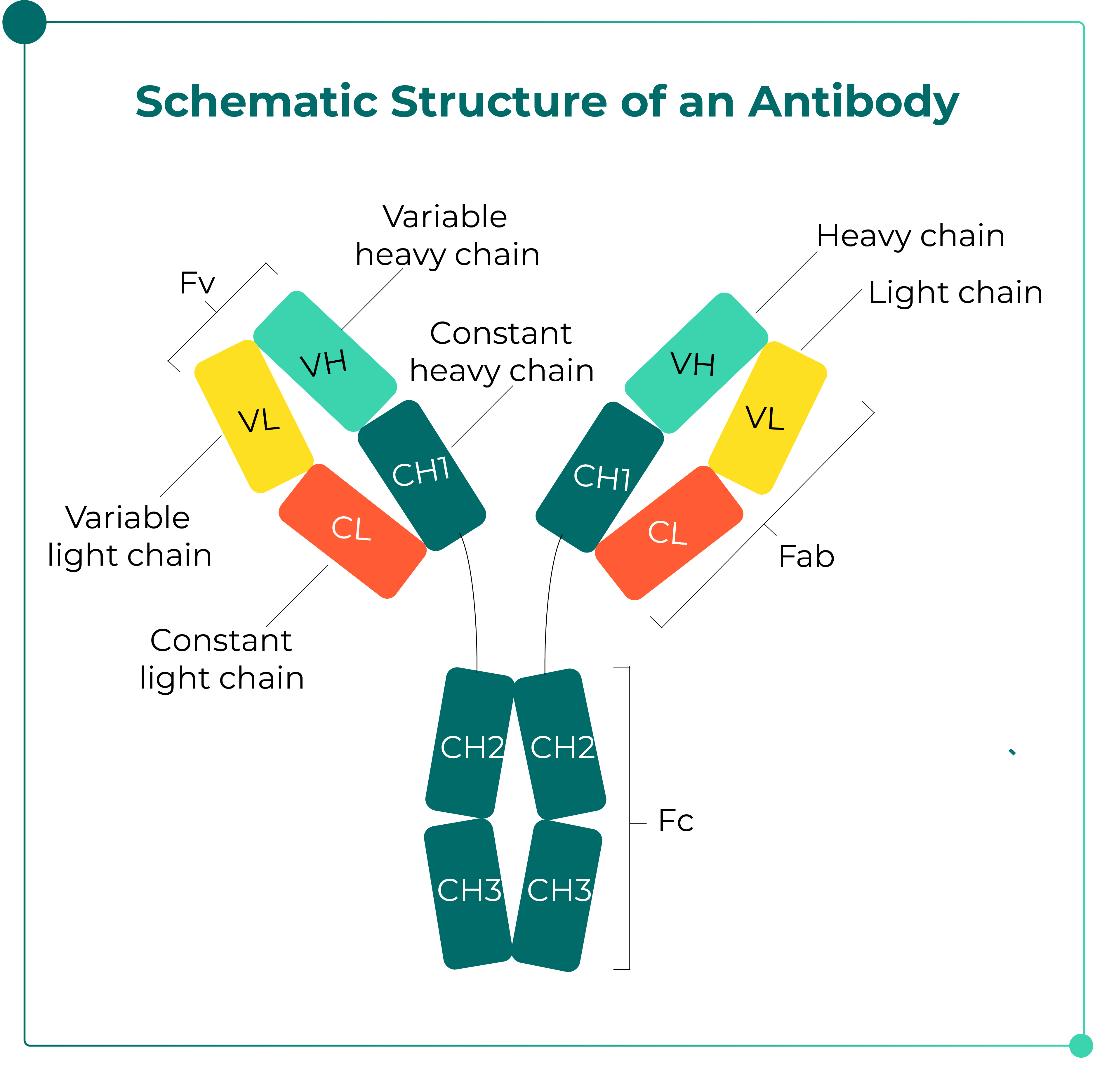
Fig. 1. Schematic Structure of an Antibody
Light and heavy chains: Each light chain consists of approximately 214 amino acids with a molecular weight of about 24 kDa, and is classified into two types: κ light chain and λ light chain. By contrast, each heavy chain contains approximately 450–550 amino acids, with a molecular weight of 55–75 kDa, and typically includes glycosylation modifications.
Domain classification: Each light and heavy chain can be further divided into a variable region (V region) and a constant region (C region). The V region is located at the N-terminus and is responsible for specific antigen recognition and binding, while the C region is located at the C-terminus, which is relatively conserved in sequence, and mediates immune effector functions.
Antigen-binding site: Within the V region, highly variable amino acid segments known as hypervariable regions (HVRs), also referred to as complementarity-determining regions (CDRs). Each light chain and heavy chain contains three CDRs, which together constitute the antigen-binding sites and determine antibody specificity.
Domains and hinge region: Antibody molecules fold into globular domains via intrachain disulfide bonds, with each domain comprising approximately 110 amino acids. In IgG, IgA, and IgD, each heavy chain contains four domains (one V domain and three C domains), whereas in IgM and IgE, each heavy chain contains five domains (one V domain and four C domains). The hinge region, located between CH1 and CH2 of the heavy chains in IgG, IgA, and IgD, is rich in proline residues and provides structural flexibility, enabling more effective antigen binding. However, IgM and IgE lack a classical hinge region.
Based on differences in the amino acid composition and arrangement of the heavy chain constant regions, antibodies are classified into five major isotypes: IgG, IgM, IgA, IgD, and IgE, corresponding to γ, μ, α, δ, and ε heavy chains, respectively.
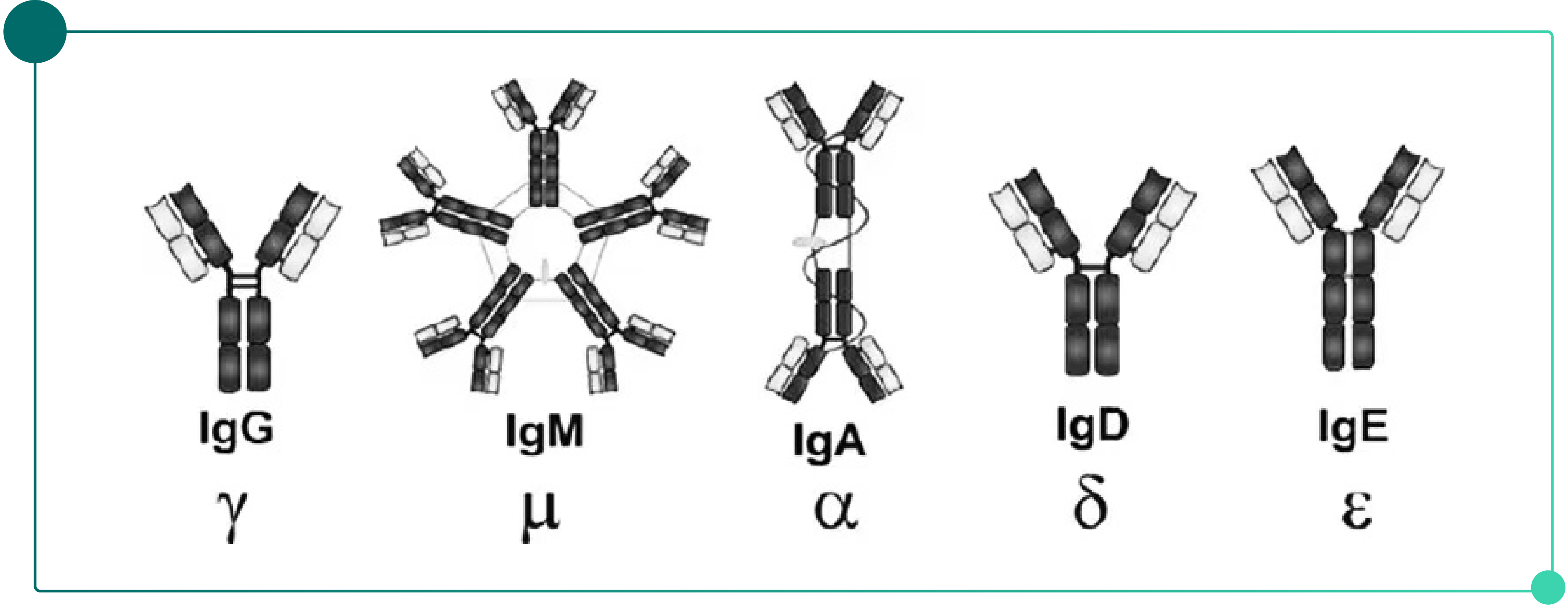
Fig. 2. Classification of Antibodies
IgG — the dominant format for therapeutic antibodies: IgG is the most abundant antibody in serum and the most commonly used molecular format in antibody therapeutics. Based on differences in hinge region structure and the position of interchain disulfide bonds, human IgG can be further classified into four subclasses: IgG1, IgG2, IgG3, and IgG4. Among these, IgG1 serves as the preferred backbone for vast majority of antibody drugs due to its well-balanced effector functions and stability.
Distinct structures of IgM and IgA: IgM exists predominantly as a pentamer containing a J chain, giving it the largest molecular size among immunoglobulins. IgA can exist as a monomer or form a dimer via a J chain, and is mainly distributed on mucosal surfaces.
Light chain classification: Based on differences in the constant region of the light chain, antibodies are classified into κ (kappa) and λ (lambda) types. Accordingly, immunoglobulins can also be categorized into κ-type and λ-type.
Based on structural characteristics, antibody therapeutics can be broadly classified into four major categories:

Fig.3. Major Types of Antibody Therapeutics
1) Antibody fragments (Fc-free):
These molecules lack the Fc region and function primarily through antigen-binding domains. They typically have a low molecular weight and high tissue penetration. As shown in Fig. 3 (left, examples include Fab fragments, F(ab')₂ fragments, single-chain variable fragments (scFv), diavalent scFv (di-scFv), and single-domain antibodies such as sdAb/VHH (nanobodies).
2) Monoclonal antibody (mAb):
Monoclonal antibodies represent the foundational format of antibody therapeutics. They are homogeneous antibodies produced by a single B cell clone and typically adopt the classical IgG structure, consisting of two heavy chains and two light chains, with an intact Fc region.
3) Bispecific Antibody,BsAb / Multispecific Antibody,MsAb
These engineered antibodies are designed to simultaneously recognize two or more distinct targets within a single molecule, enabling more complex biological functions. Examples include IgG-like BsAbs bispecific antibodies, non-IgG-like bispecific formats, trispecific antibodies (TsAbs), etc.
4) ADC/XDC (antibody–drug conjugates):
Antibody–drug conjugates (ADCs) consist of three components: an IgG antibody, a linker, and a cytotoxic drug (payload). The antibody specifically binds to target antigens on the surface of tumor cells, and after internalization, the cytotoxic drug is released, achieving precise killing while reducing systemic toxicity. For a detailed discussion of ADC downstream purification strategies, see Downstream Purification Strategies for Antibody–Drug Conjugates.
Expansion of the XDC modalities: With technological advancements, XDC (X-Drug Conjugates) has emerged as an umbrella term for a broader range of conjugated therapeutics, including RDCs (radionuclide conjugates), SMDCs (small molecule drug conjugates), PDCs (peptide-drug conjugates), AOCs (antibody-oligonucleotide conjugates), VDCs (virus-like particle conjugates), ISACs (immune-stimulating antibody conjugates), and FDCs (fragment-drug conjugates).
Chromatography is the core technique for antibody purification. Based on different separation principles, it can be broadly categorized into four main types, each with its unique advantages and application scenarios. The selection of an appropriate purification method is based on the physicochemical properties of the target protein and the associated impurities.
|
Differences Between Target Protein and Impurities |
Separation Technique |
|
Specific Functional Group |
Affinity Chromatography (AC) |
|
Isoelectric Point (pI) |
Ion Exchange Chromatography (IEX); Mixed-mode Chromatography (MMC/MMA) |
|
Hydrophobicity |
Hydrophobic Interaction Chromatography (HIC); Mixed-mode Chromatography (MMC/MMA) |
|
Molecular Size |
Gel Filtration (GF) |
1) Affinity Chromatography (AC) — Specific Interactions
Principle: Affinity chromatography is a separation technique based on specific interactions between biomolecules, such as enzyme–substrate, receptor–ligand, and antibody–antigen binding. These interactions are both highly specific and reversible, enabling selective binding and subsequent elution to achieve protein purification.
Due to its high selectivity, affinity chromatography can rapidly capture target proteins from complex mixtures, tipically achieving purity above 90% in a single step. It is straightforward to operate and well-suited for use as the primary capture step in antibody purification processes.
2) Ion Exchange Chromatography (IEX) — Isoelectric Point (pI)
Principle: Ion exchange chromatography separates biomolecules based on differences in the type and magnitude of their net charge. Most biomolecules contain acidic or basic groups, and their charge properties can be modulated by adjusting the pH of the buffer.
After binding to oppositely charged resins—either anion exchange or cation exchange—proteins can be eluted by altering the ionic strength or pH of the mobile phase. Weakly bound molecules are eluted first, followed by strongly bound ones, thereby achieving separation and purification.
Based on the charge of the functional ligands, ion exchange chromatography is classified into anion exchange chromatography (AEX) and cation exchange chromatography (CEX).
3) Hydrophobic Interaction Chromatography (HIC) — Hydrophobicity
Principle: Hydrophobic interaction chromatography is a widely used technique for separating proteins and other biomolecules based on differences in surface hydrophobicity.
The key to selecting a HIC resin lies in choosing a ligand with appropriate hydrophobicity Proteins with higher hydrophobicity generally require resins with weaker hydrophobic ligands, whereas less hydrophobic proteins may require stronger hydrophobic ligands to achieve effective separation.
4) Gel Filtration (GF) — Molecular Size
Principle: Gel filtration chromatography separates biomolecules based on differences in their size and shape. The gel filtration resin consists of a porous matrix, where smaller molecules penetrate deeper into the pores and therefore have longer residence time, while larger molecules are excluded and elute earlier. Each chromatographic resin has a defined pore size distribution. The key to selecting an appropriate SEC resin is choosing a suitable separation range, followed by consideration of the resin's mechanical properties and scalability.
Antibody purification typically consists of a capture step (e.g., Protein A/G/L affinity chromatography) followed by polishing steps such as ion exchange, gel filtration, and hydrophobic interaction chromatography, ultimately yielding a highly purified antibody product.
A typical antibody purification process includes the following key unit operations:
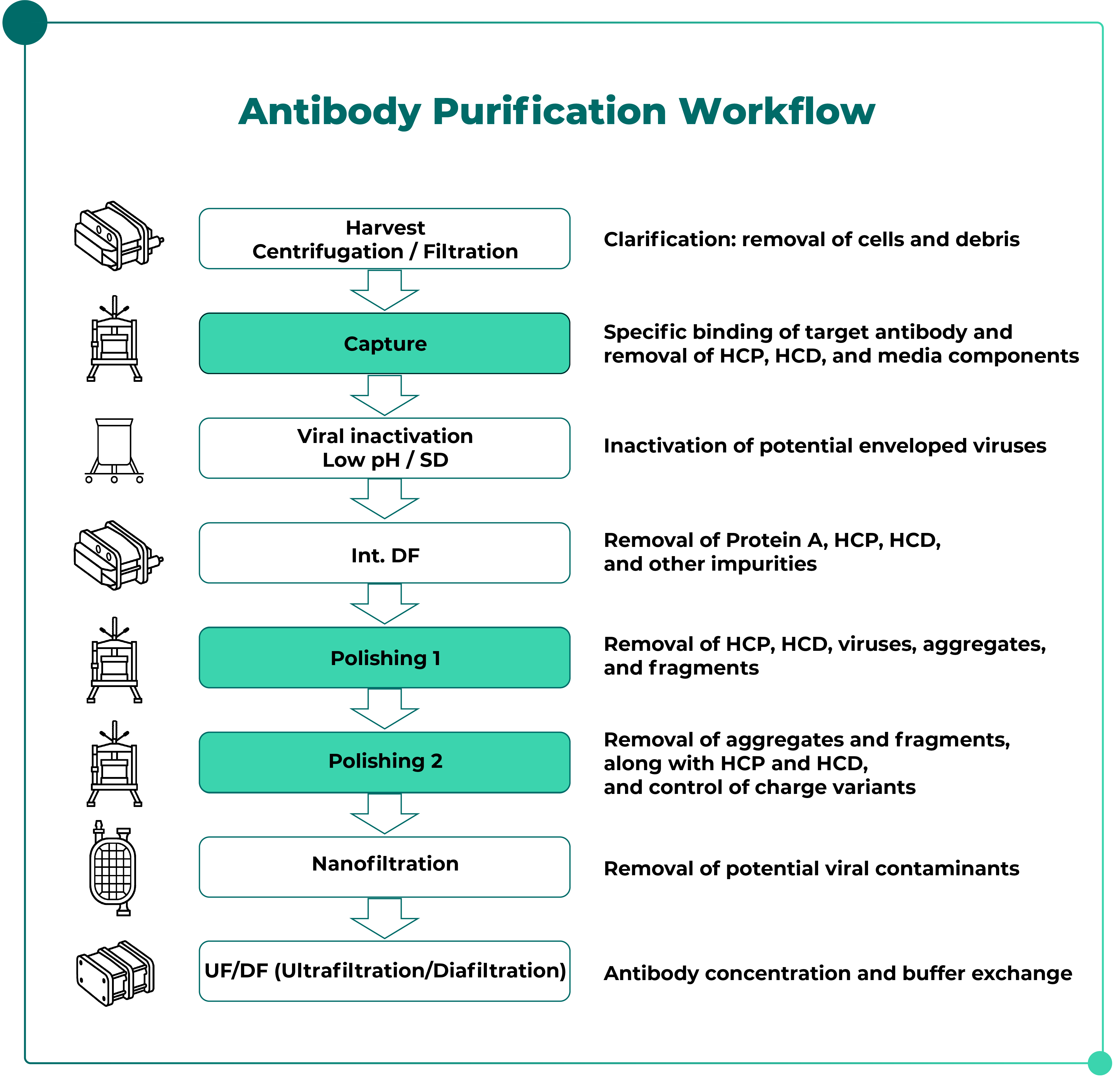
Fig. 4. Typical Antibody Purification Workflow
The capture step in antibody purification is typically achieved using antibody affinity chromatography. This technique leverages specific interactions between biomolecules to enable highly selective separation.
The most commonly used ligand is Protein A-a membrane protein derived from Staphylococcus aureus, which specifically binds to the Fc region of antibodies, enabling efficient one-step capture and high-purity purification.
1) Protein A Affinity Chromatography
Native Protein A contains five homologous IgG-binding domains (E, D, A, B, and C), but the full-length molecule also includes regions responsible for non-specific binding. Therefore, most modern commercial resins employ genetically engineered Protein A ligands to significantly reduce non-specific binding and enhance alkaline stability. For a comprehensive overview of resin options and selection criteria, refer to Everything You Need to Know About Protein A Affinity Resins.
In addition, Protein A domains can also interact with the VH region of Fab fragments. These interactions involve multiple forces, including hydrophobic interactions, hydrogen bonding, and salt bridges.
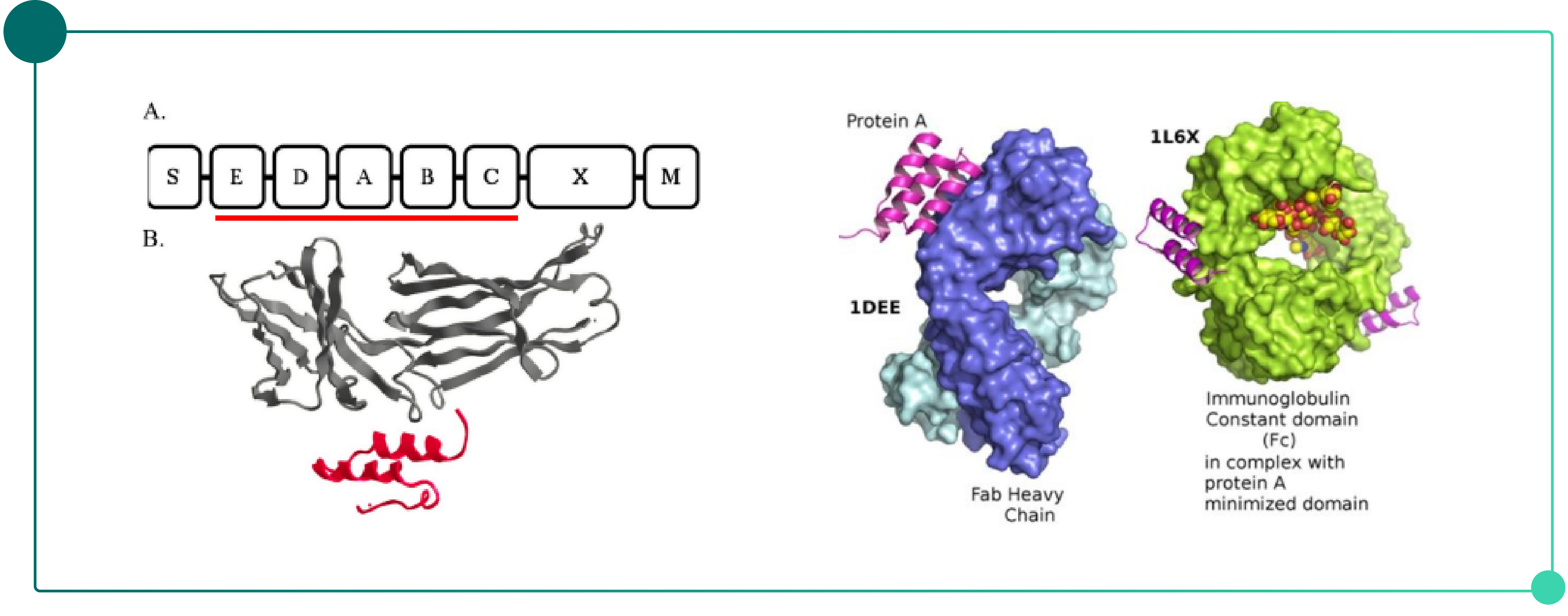
Fig. 5. Binding Mechanism of Protein A Affinity Chromatography
2) Other Antibody Affinity Chromatography Approaches
For specific molecules such as Fab fragments, IgM, and other antibody fragments, alternative affinity ligands can be employed for selective separation. For example, Protein L (which binds to κ chain) or IgM-specific affinity resins can be used.
Following the capture step—typically affinity chromatography—antibody purity usually exceeds 90%, with significantly reduced levels of host cell proteins (HCP) and host cell DNA (HCD). However, further removal is still required in subsequent steps. Furthermore, product-related impurities such as aggregates, fragments, and charge variants, as well as process-related impurities introduced during affinity capture (e.g., residual Protein A), must be eliminated through further polishing steps, such as IEX, MMC/MMA, HIC and GF.
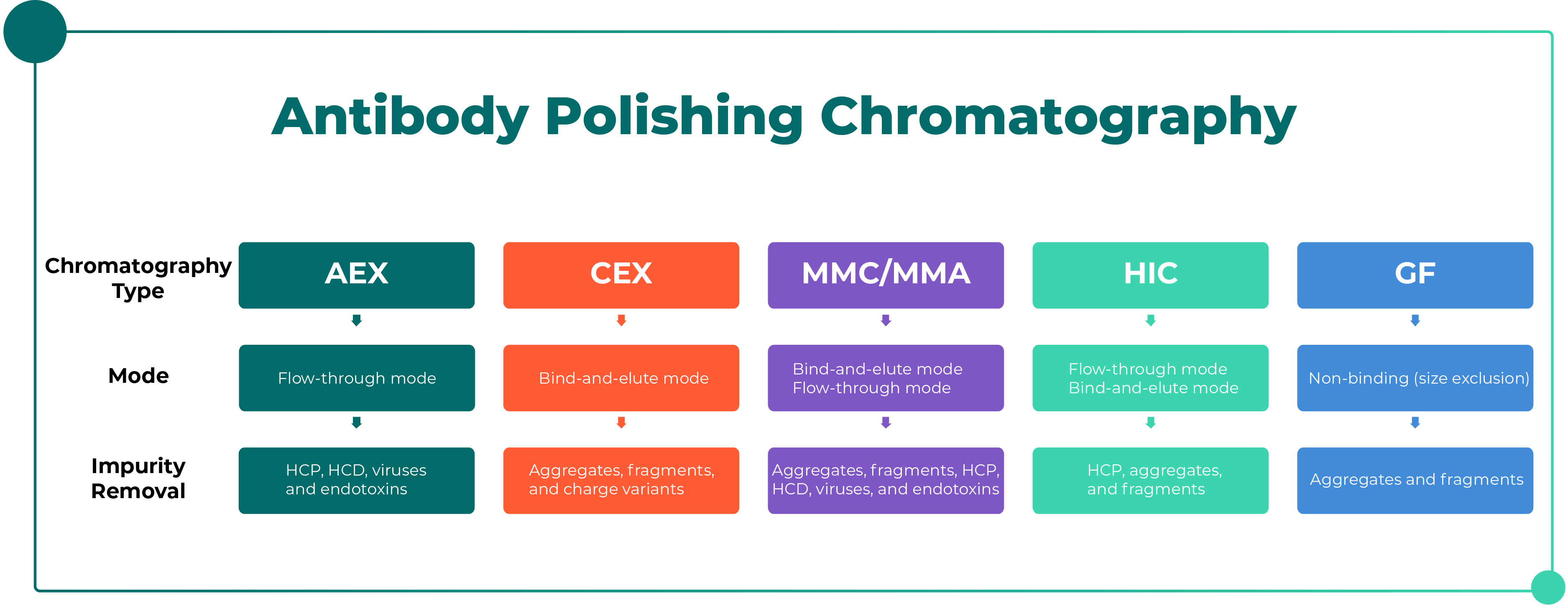
1) Anion Exchange Chromatography (AEX) — Flow-through Mode
This is the most commonly used operation mode in antibody polishing. It primarily removes process-related impurities such as HCP, HCD, Virus, and is often considered an intermediate purification step.
2) Cation Exchange Chromatography (CEX) — Bind-and-Elute Mode
Cation exchange chromatography is typically operated in bind-and-elute mode, enabling the removal of both process-related impurities (HCP, HCD) and product-related impurities such as aggregates, fragments, and charge variants.
3) MMC/MMA — Bind-and-Elute or Flow-through Mode
Mixed-mode chromatography combines ion exchange and hydrophobic interaction mechanisms. It can be operated in either bind-and-elute or flow-through mode to remove product-related impurities (e.g., aggregates and fragments) and/or process-related impurities such as HCP, HCD, endotoxins, and viruses.
4) HIC — Flow-through / Bind-and-Elute Mode
When ion exchange chromatography is insufficient for aggregate removal, HIC can serve as a complementary or alternative approach. It is commonly operated in either flow-through or bind-and-elute mode to remove impurities such as HCP, aggregates, and fragments.
5) GF — Flow-through / Bind-and-Elute Mode
Gel filtration is a non-binding technique that separates molecules based on size differences between the target protein and impurities. It is simple to operate and often used as a final polishing step or for buffer exchange/desalting. However, due to its low sample loading capacity, it is generally avoided in large-scale manufacturing.
· Resin inactivation: Ligands (e.g., Ni²⁺, antibodies) lose activity due to repeated use, improper cleaning, or unsuitable storage conditions.
· Sample-related issues:
1) High sample viscosity
2) Presence of high concentrations of competitive agents (e.g., imidazole in the sample when applying IMAC; high concentration of DTT can reduce metal ions)
· Inappropriate buffer conditions:
1) pH value: outside the stable range of the protein or ligand
2) Chelating agents: in IMAC (e.g., Ni columns), specific buffer components such as EDTA can strip metal ions from the resin
· Excessive flow rate: Insufficient contact time between the protein and the ligand reduces binding efficiency
· Tag-related issues (for tagged protein purification, e.g., His-tag):
1) Tag protein is not expressed along with target protein or incompletely expressed
2) Tag protein is masked (e.g., due to inclusion body formation or higher-order protein structure)
The properties of pigments are complex, and CIP protocols can be developed based on resin tolerance. NaOH is the most commonly used cleaning agent for CIP. In addition, high-salt solutions, organic solvents (e.g., 20% ethanol or 30% isopropanol), urea, guanidine hydrochloride, or acidic solutions are also common cleaning options. Mixed cleaning agents or other targeted cleaning approaches may be applied as needed. If the type of pigment is known, specific cleaning methods can be selected based on its chemical properties.
· Adjust salt concentration or optimize the pH of the sample
· Introduce additional wash steps (e.g., high-salt or organic solvent washes)
· Optimize the elution pH
This may be due to an inappropriate elution gradient, and a shallower gradient may improve resolution. Other factors may include an excessively high flow rate or protein aggregation during elution. Alternatively, switching to a resin with higher resolution may also improve separation performance.
The sample may not bind due to excessively high salt concentration, inappropriate pH conditions, the presence of charged components in the buffer (e.g., detergents) or decreased column performance. These issues can be addressed by evaluating column efficiency, optimizing sample loading conditions, etc.
It is recommended to increase the salt concentration in both the equilibration buffer and the sample, or to use salts with stronger salting-out effects. If there is still no improvement, alternative ligands or hydrophobic resins with higher ligand density can be evaluated.
Antibody purification is a process that balances resolution, binding capacity, processing speed, recovery against cost. Starting from the structural characteristics and classification of antibodies, a thorough understanding of the principles and application scenarios of different chromatographic techniques, along with the rational design of capture and polishing strategies, is key to establishing efficient and robust purification process.
As antibody formats become increasingly diversify (e.g., BsAbs ADCs, AOCs, etc.), conventional platform purification processes face increasing challenges. Greater variability in Protein A binding, increased sensitivity of molecular stability, and closer similarity between impurities and target molecules all place higher demands on ligand selection, elution conditions, and cleaning strategies. The development of affinity resins that combine high binding capacity, high selectivity, and mild elution conditions will be a key direction in antibody purification process development.


Jul
21

Jul
16
Jun
26
.png)


.png)
.png)
.png)

.png)